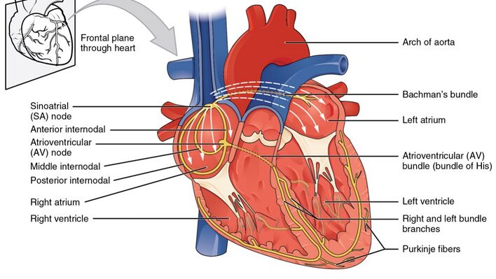
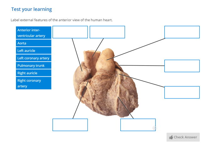
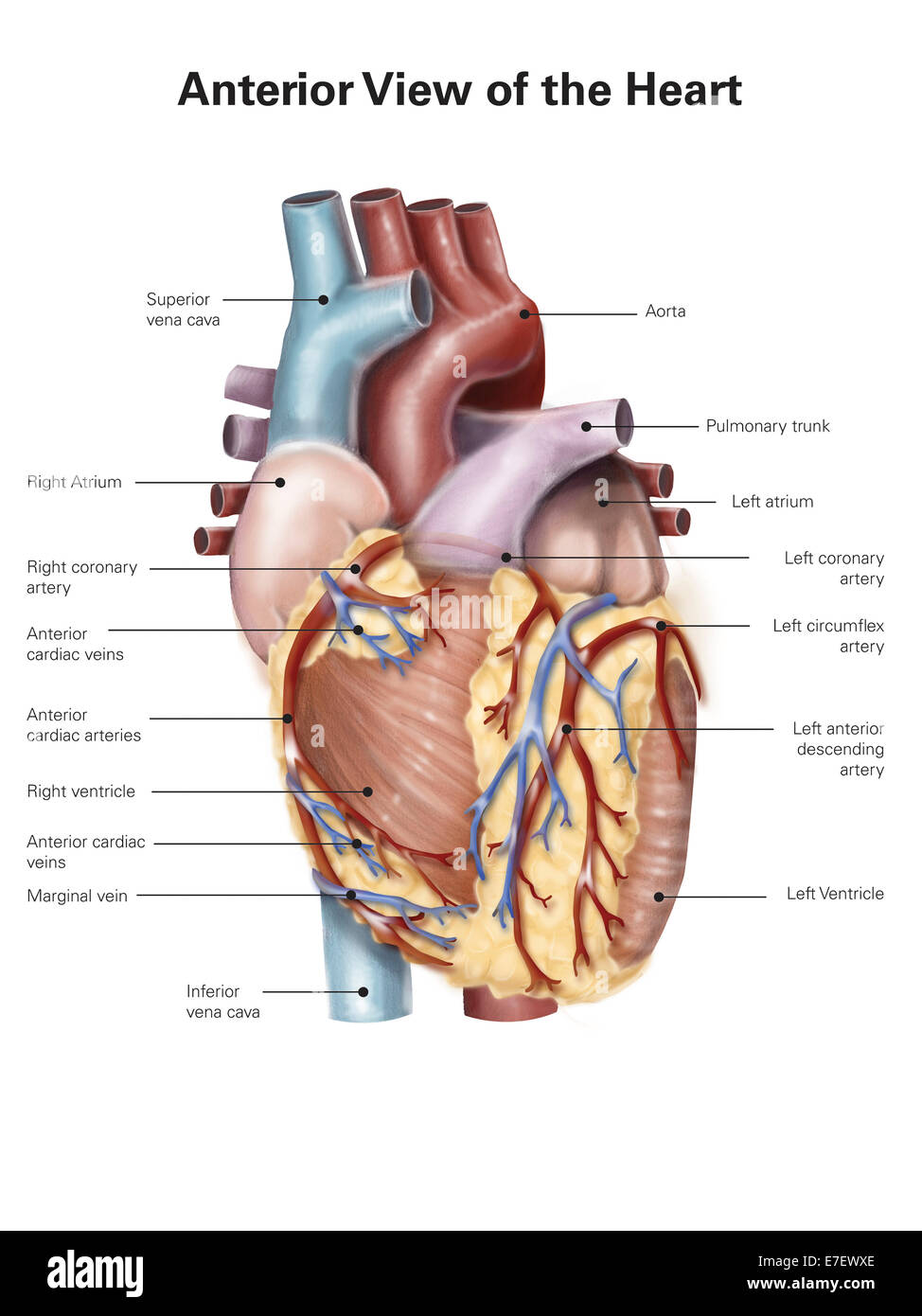
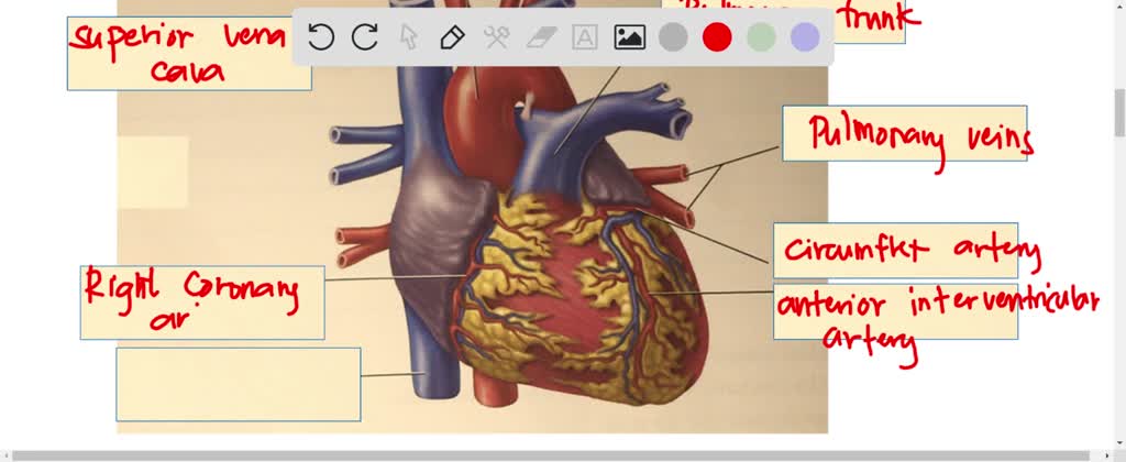
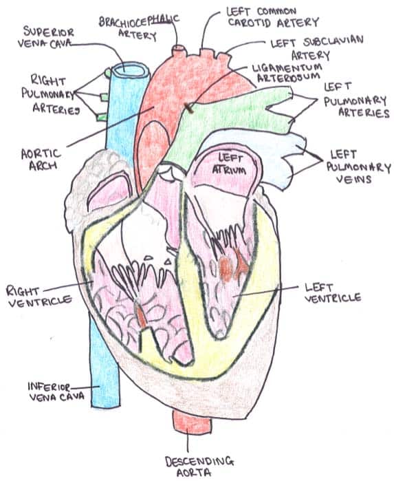
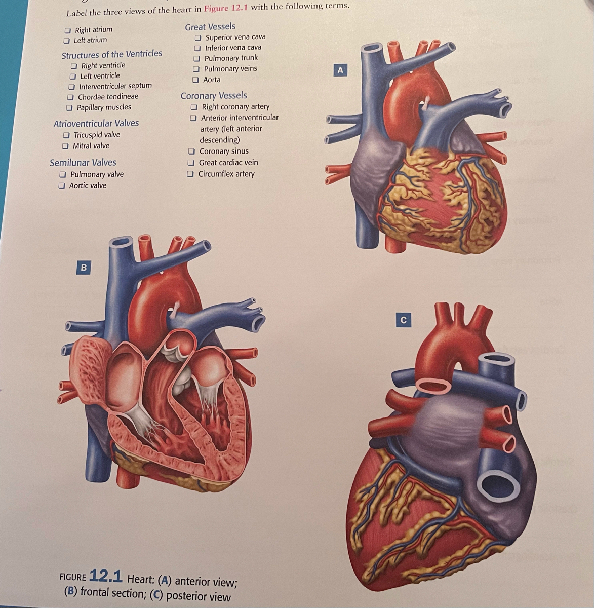
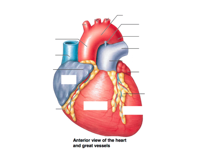
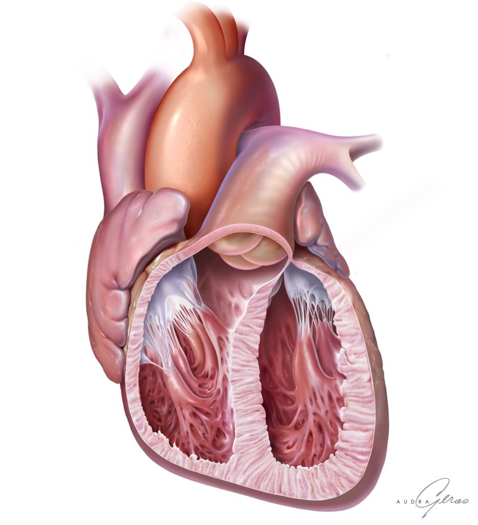
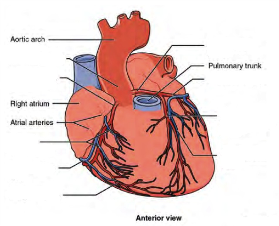
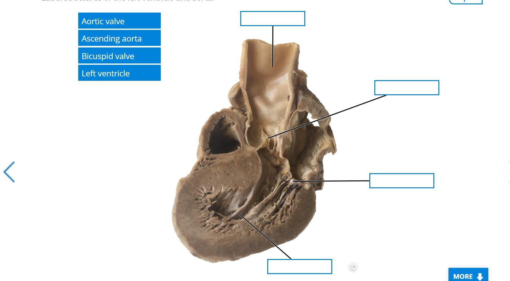
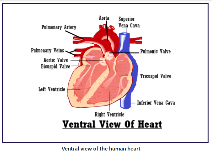
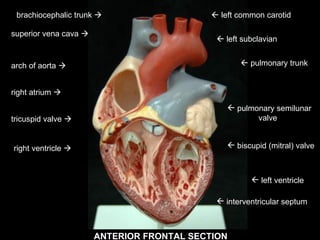
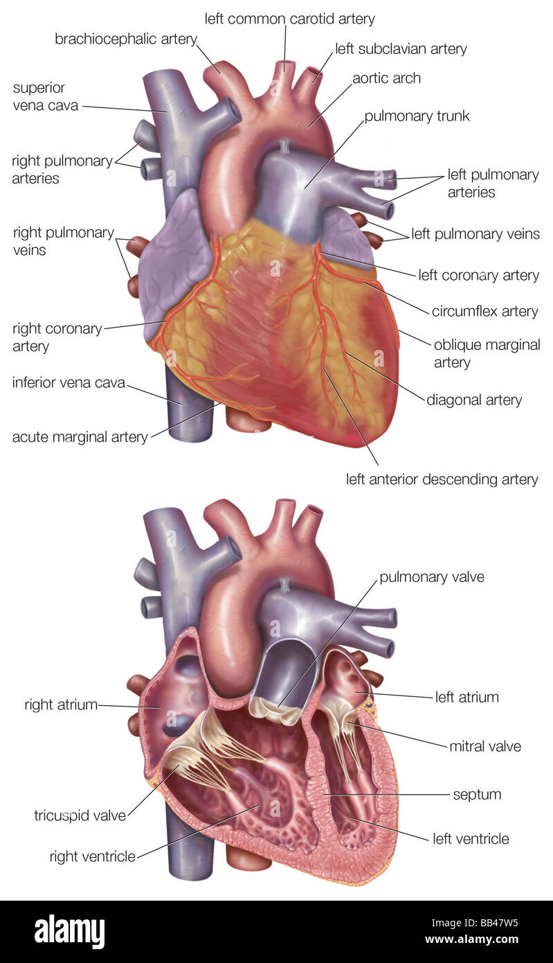
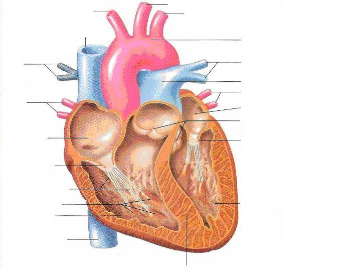
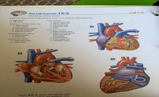
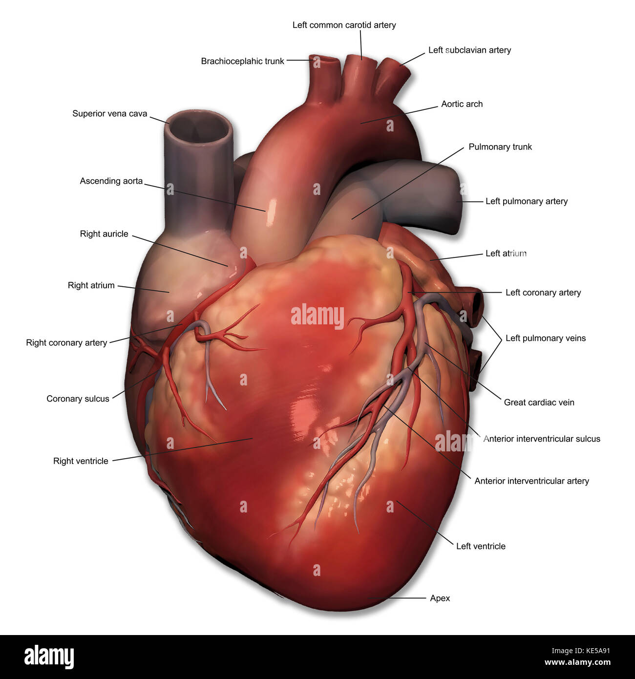
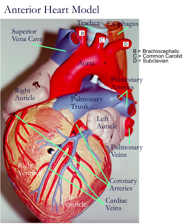
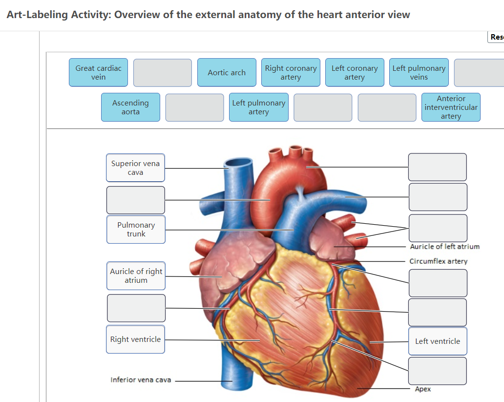
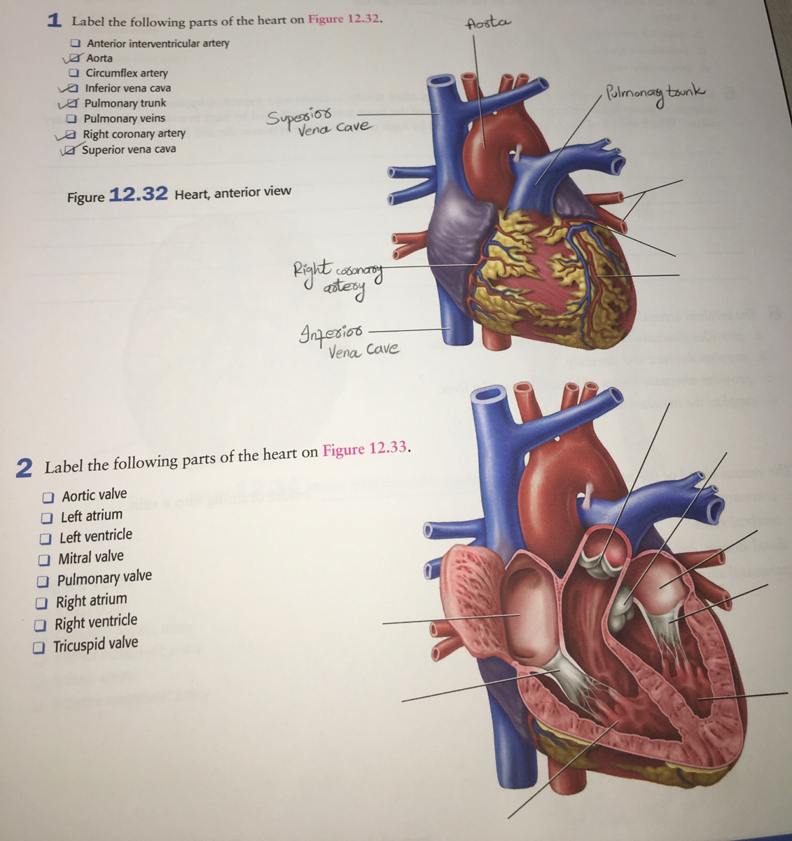
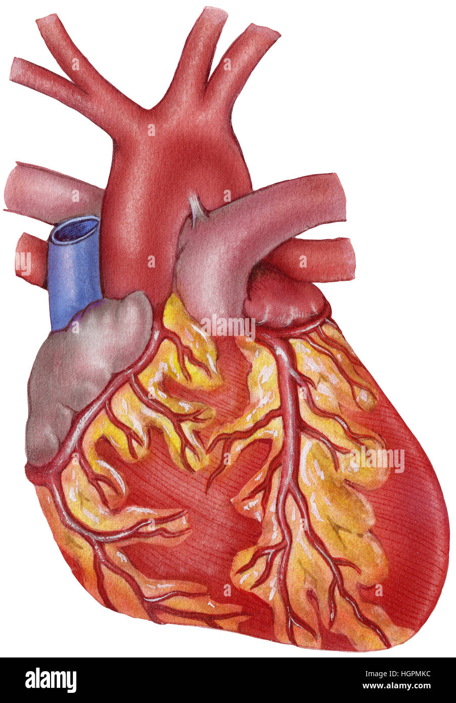
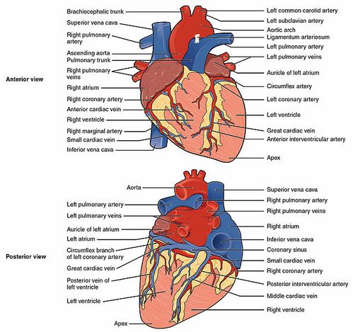
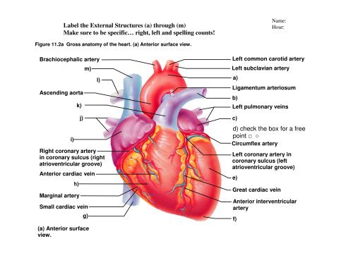
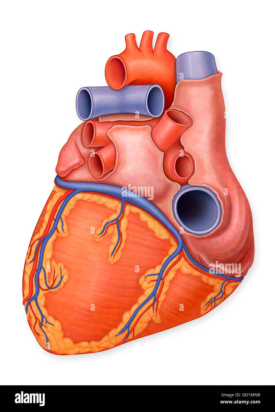
43 label the anterior view of the human heart
Heart: illustrated anatomy | e-Anatomy - IMAIOS This interactive atlas of human heart anatomy is based on medical illustrations and cadaver photography. The user can show or hide the anatomical labels which provide a useful tool to create illustrations perfectly adapted for teaching. ... Left ventricle, Left atrium, Anterior papillary muscle Pericardium : Pericardial cavity, Transverse ... Lab 44- Heart Structure Flashcards | Quizlet Label the anterior heart structures by clicking and dragging the labels to the correct location. Bottom (2): 1. Left ventricle 2. Apex Right side: 1.)Ligamentrum 2.)left pulmonary artery 3.)Pulmonary trunk 4.)Left pulmonary veins 5.)Auricle of left atrium 6.)Grat cardiac vein 7.)Anterior interventricular artery
Heart Labeling anterior view Diagram | Quizlet Heart Labeling anterior view Diagram | Quizlet Heart Labeling anterior view 5.0 (3 reviews) + − Learn Test Match Created by Meghan12th Terms in this set (26) Term brachiocephalic trunk Location Term left common carotid artery Location Term superior vena cava Location Term aortic arch Location Term liigamentum arteriosum Location Term

Label the anterior view of the human heart
Heart Lab Flashcards | Quizlet Label the chambers and valves seen in an anterior view of the heart. Fill in the blanks with the appropriate words to describe blood flow from the heart. Then place the sentences in order to form a coherent paragraph. Label the coronary arteries on the posterior surface of the heart. Label the cardiac veins on the posterior surface of the heart Anterior External View of Heart Labeling Diagram | Quizlet Anterior External View of Heart Labeling Diagram | Quizlet Anterior External View of Heart Labeling 5.0 (2 reviews) Learn Test Match Created by randajsmith Teacher Terms in this set (14) Superior Vena Cava ... Term Superior Vena Cava (SVC) Location Term Right Pulmonary Artery Location Term Right Pulmonary Veins Location Term Right Atrium Location Lab Report 38 Figures 38.1 38.2 and 38.3.pdf - Figure 38.1- Label this ... Figure 38.2- Label this posterior view of the human heart. 1.) Aorta 2.) Superior vena cava3.) Aortic valve4.) Right atrium5.) Tricuspid valve6.) Chordae tendness7.) Inferior vena cava8.) Left pulmonary artery 9.) Pulmonary trunk10.) Left pulmonary veins11.) Left atrium12.) Pulmonary valve 13.) Mitral valve14.) Papillary muscle15.)
Label the anterior view of the human heart. ch. 13 Homework Flashcards | Quizlet Study with Quizlet and memorize flashcards containing terms like Label the components of the cardiac conduction system., Label the veins of the lower limb., Place the following structures in order through which the blood flows. and more. ... Label the vessels seen in the anterior view of the frontal section of the heart. 1. The tricuspid valve ... Heart: Anatomy and Function - Cleveland Clinic Left anterior descending artery (LAD): Supplies blood to the front and bottom of the left ventricle and the front of the septum. Right coronary artery (RCA): Supplies blood to the right atrium, right ventricle, bottom portion of the left ventricle and back of the septum. Electrical conduction system Human Heart (Anatomy): Diagram, Function, Chambers, Location in Body An echocardiogram provides direct viewing of any problems with the heart muscle's pumping ability and heart valves. Cardiac stress test: By using a treadmill or medicines, the heart is... Figure 35.3 Frontal Section of Human Heart Diagram | Quizlet Term Aortic Valve Location Term Mitral (bicuspid) Atrioventricular Valve Location Term Right Atrium Location Term Tricuspid Atrioventricular Valve Location Term Chordae Tendineae Location Term Inferior Vena Cava Location Term Papillary Muscle Location Term Interventricular Septum Location Term Left Ventricle Location Term Right Ventricle Location
Figure 35.1 Heart (ventral view) Diagram | Quizlet Start studying Figure 35.1 Heart (ventral view). Learn vocabulary, terms, and more with flashcards, games, and other study tools. ... Figure 35.3 Frontal Section of Human Heart. 17 terms. Diagram. THagge Teacher. Figure 35.2 Heart (dorsal view) 10 terms. Diagram. THagge Teacher. Figure 35.4 Heart Model (anterior view) 11 terms. Diagram. THagge ... Heart anatomy: Structure, valves, coronary vessels | Kenhub The heart has five surfaces: base (posterior), diaphragmatic (inferior), sternocostal (anterior), and left and right pulmonary surfaces. It also has several margins: right, left, superior, and inferior: The right margin is the small section of the right atrium that extends between the superior and inferior vena cava . Chapter 22 Heart Flashcards | Quizlet Label the valves in an anterior view of the heart. Label the coronary arteries in an anterior view of the heart. Label the order that blood flows through in the heart, using the arrows as guides. Label the components of the heart wall. Label the components of the heart as seen from a posterior view. Label the major coronary veins. Lab Report 38 Figures 38.1 38.2 and 38.3.pdf - Figure 38.1- Label this ... Figure 38.2- Label this posterior view of the human heart. 1.) Aorta 2.) Superior vena cava3.) Aortic valve4.) Right atrium5.) Tricuspid valve6.) Chordae tendness7.) Inferior vena cava8.) Left pulmonary artery 9.) Pulmonary trunk10.) Left pulmonary veins11.) Left atrium12.) Pulmonary valve 13.) Mitral valve14.) Papillary muscle15.)
Anterior External View of Heart Labeling Diagram | Quizlet Anterior External View of Heart Labeling Diagram | Quizlet Anterior External View of Heart Labeling 5.0 (2 reviews) Learn Test Match Created by randajsmith Teacher Terms in this set (14) Superior Vena Cava ... Term Superior Vena Cava (SVC) Location Term Right Pulmonary Artery Location Term Right Pulmonary Veins Location Term Right Atrium Location Heart Lab Flashcards | Quizlet Label the chambers and valves seen in an anterior view of the heart. Fill in the blanks with the appropriate words to describe blood flow from the heart. Then place the sentences in order to form a coherent paragraph. Label the coronary arteries on the posterior surface of the heart. Label the cardiac veins on the posterior surface of the heart








Post a Comment for "43 label the anterior view of the human heart"