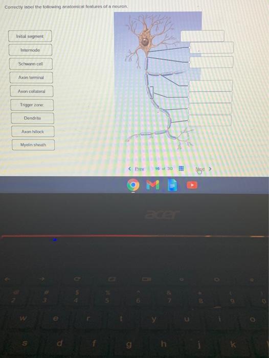
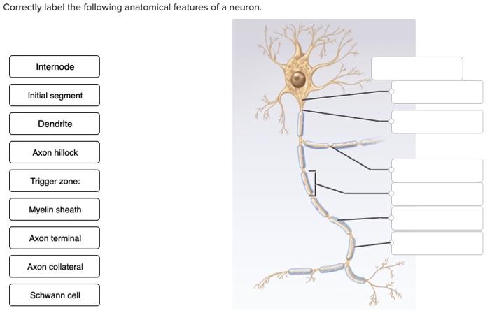
41 correctly label the following anatomical features of a neuron
Multipolar Neurons - Structure, functions and diagram | GetBodySmart Multipolar neurons have three or more processes attached to the cell bodies. 1. 2. One process serves as the axon, which conducts electrochemical impulses ( action potentials) between cells. 1. 2. The remaining processes are dendrites. Together, the cell body and dendrites form the receptive zone of multipolar neurons. 1. Parts of a neuron labeled and neuron structure | GetBodySmart Neurons (nerve cells) are the functional units of the nervous system. Even though they vary in size and shape, most have structural characteristics similar to the spinal cord neuron shown to left. Neurons have at their core an expanded area of cytoplasm called the cell body (soma or perikaryon). 1. 2.
Neuron | Journal | ScienceDirect.com by Elsevier Neuron has established itself as one of the most influential and relied upon journals in the field of neuroscience. The editors embrace interdisciplinary strategies which integrate biophysical, cellular, developmental, and molecular approaches with a systems approach to sensory, motor, and …. View full aims & scope.

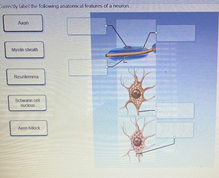
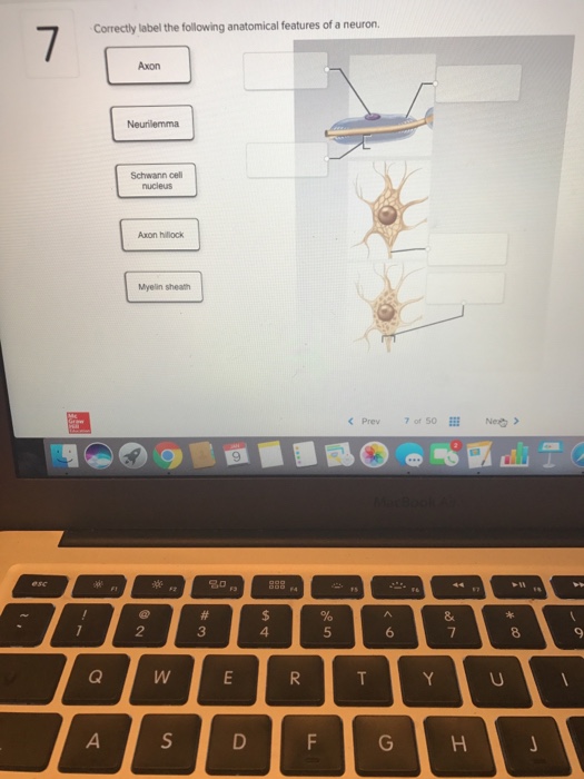
Correctly label the following anatomical features of a neuron
Neuron Cell Body - Structure and Functions - GetBodySmart Neuron cell bodies basically have the same cytoplasmic components as other types of secretory cells. The cell's large nucleus and nucleolus are the most prominent cell body structures. 1. 2. Group of free ribosomes and and numerous stack of ribosome studded rough endoplasmic reticulum (REP) surround the nucleus. 1. 2. 3. Classify each of the compounds as soluble or not soluble:...open 8 Answer of Classify each of the compounds as soluble or not soluble: cobalt(II) hydroxide iron(II) acetate ammonium sulfide Correctly Label The Following Anatomical Parts Of Osseous Tissue Adjust Credit For All Students. 1 three layers tunics that form its wall 2 optical components that admit and focus light and 3 neural components the retina and optic nerve. 100% (42 ratings) transcribed image text: Correctly label the following anatomical parts of. Related posts: Label A Label B Label C Label D Historians Label An Event As A ...
Correctly label the following anatomical features of a neuron. Nervous system: Structure, function and diagram | Kenhub Neurons, or nerve cell, are the main structural and functional units of the nervous system. Every neuron consists of a body (soma) and a number of processes (neurites). The nerve cell body contains the cellular organelles and is where neural impulses ( action potentials) are generated. The processes stem from the body, they connect neurons with ... Which Of The Following Is Not A Function Of Neurons 34 Correctly Label The Following Anatomical Features Of A Neuron from ugarevwesi.blogspot.com. If you think about the roles of the three classes of neurons, you can make the generalization that all neurons have three basic functions. This quiz on the neural structure will help you when you are studying for your exam in a & p. Correctly Label Tooth Anatomical Of The A Features Search: Correctly Label The Anatomical Features Of A Tooth. Look here for the answers!!! Pick a Bone! Skull: anterior lateral posterior The cranium (skull) is the skeletal structure of the head that supports the face and protects the brain Question: Correctly label the anatomical features of a tooth Gingiva Neck Pulp cavity Dentin Cementum Root Root canal Periodontal ligament Bone Gingival ... 3 Correctly label the following anatomical features of the...open 8 Answer of 3 Correctly label the following anatomical features of the lymph node Efterent lymphatic vessel eBook Germinal center Cortical sinus Lymphatic nodule...
Spinal cord: Anatomy, structure, tracts and function | Kenhub Anatomy. The spinal cord is part of the central nervous system (CNS). It is situated inside the vertebral canal of the vertebral column. During development, there's a disproportion between spinal cord growth and vertebral column growth. The spinal cord finishes growing at the age of 4, while the vertebral column finishes growing at age 14-18. Action potential - Definition, Steps, Phases | Kenhub An action potential is defined as a sudden, fast, transitory, and propagating change of the resting membrane potential. Only neurons and muscle cells are capable of generating an action potential; that property is called the excitability. This article will discuss the definition, steps and phases of the action potential. 9+ which of the following is not part of a neuron most standard 1.Which of the following is not part of a neuron? a. cell body b. axon c … 2.Which of the following are NOT part of a neuron? a) synapse b) axon … 3.Which of the following is not a part of the neuron? - Byju's; 4.Which of the following is not part of a neuron? - Toppr; 5.Solved Which of the following is not part of a neuron? | Chegg.com Anatomy Homework Help & Textbook Solutions - Quesba 3 Correctly label the following anatomical features of the lymph node Efterent lymphatic vessel... 3 Correctly label the following anatomical features of the lymph node Efterent lymphatic vessel eBook Germinal center Cortical sinus Lymphatic nodule Cortex Medulla Subcapsular sinus Trabecula Previous question.
Correctly label the following anatomical features of the...open 8 Answer of Correctly label the following anatomical features of the thoracic cavity. (Not all words will be used.) Left lung Apex of heart Parietal pleura... Neuromuscular junction: Parts, structure and steps | Kenhub The presence of the synaptic cleft between the synaptic end bulb of the neuron and the motor end plate of the muscle fiber, means that the electrical signal or action potential, arriving from the central nervous system, needs to somehow transverse (cross) this space.The neuromuscular junction accomplishes this by turning the electrical signal from the nervous system into a chemical signal that ... Correctly Label The Following Anatomical Parts Of Osseous Tissue Adjust Credit For All Students. 1 three layers tunics that form its wall 2 optical components that admit and focus light and 3 neural components the retina and optic nerve. 100% (42 ratings) transcribed image text: Correctly label the following anatomical parts of. Related posts: Label A Label B Label C Label D Historians Label An Event As A ... Classify each of the compounds as soluble or not soluble:...open 8 Answer of Classify each of the compounds as soluble or not soluble: cobalt(II) hydroxide iron(II) acetate ammonium sulfide
Microtubule Dynamics Following Central and Peripheral Nervous ...
Neuron Cell Body - Structure and Functions - GetBodySmart Neuron cell bodies basically have the same cytoplasmic components as other types of secretory cells. The cell's large nucleus and nucleolus are the most prominent cell body structures. 1. 2. Group of free ribosomes and and numerous stack of ribosome studded rough endoplasmic reticulum (REP) surround the nucleus. 1. 2. 3.

Biologi Ting 4 (Form 4) | Anjung Sains Makmal 3 | Page 2

Anatomy Midterm Lecture Flashcards | Quizlet
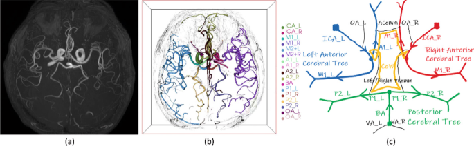
Automated Intracranial Artery Labeling Using a Graph Neural ...

8F5CA78A-C75E-4FDA-AFD9-A4516691D615.jpeg - Correctly label ...

1FA31D70-247B-4691-99E1-7C24A8844E69.jpeg - Correctly label ...
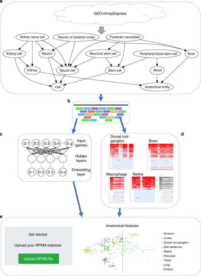
A web server for comparative analysis of single-cell RNA-seq ...

Chapter 14 Worksheet Flashcards | Quizlet

AHCDW9Notes30.pdf - 30. Award: 10.00 points Problems? Adjust ...

Anatomy Midterm Lecture Flashcards | Quizlet

BIOL 1050H Study Guide - Fall 2017, Final - Endocrine System ...

Anatomy Midterm Lecture Flashcards | Quizlet
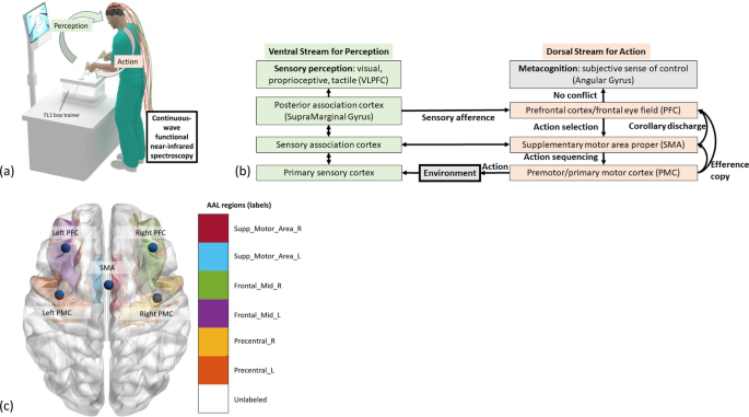
Directed information flow during laparoscopic surgical skill ...
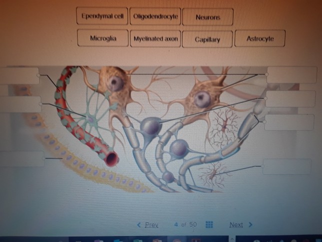
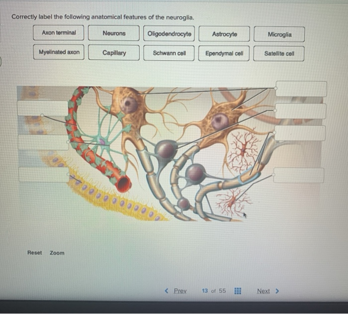
Solved Ependymal cell Oligodendrocyte Neurons | Chegg.com

BIOL 1050H Study Guide - Fall 2017, Final - Endocrine System ...
11 BAB II TINJAUAN PUSTAKA A. Lansia Lansia menurut Titus ...
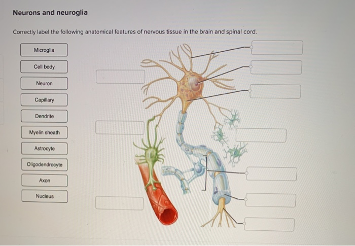
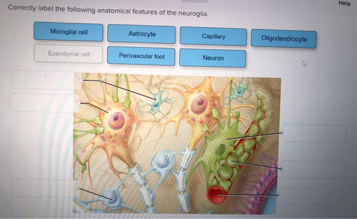
Solved Neurons and neuroglia Correctly label the following ...
Solved Correctly label the following anatomical features of ...
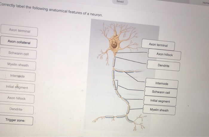
Internode Axon

AHCDW9Notes34.pdf - 34. Award: 10.00 points Problems? Adjust ...
Solved Correctly label the following anatomical features of ...

Ch 12 - Nervous System EXAM *** McGraw Flashcards | Quizlet

What Is Central Nervous System? Definition, Function & Parts
Cellular anatomy of the mouse primary motor cortex | Nature

Anatomy Midterm Lecture Flashcards | Quizlet

Anatomy Midterm Lecture Flashcards | Quizlet
Solved Correctly label the following anatomical features of ...

AHCDW8Notes6.pdf - 6. Award: 10.00 points Problems? Adjust ...

Anatomy Midterm Lecture Flashcards | Quizlet

Anatomy Midterm Lecture Flashcards | Quizlet
Solved Correctly label the following anatomical features of ...
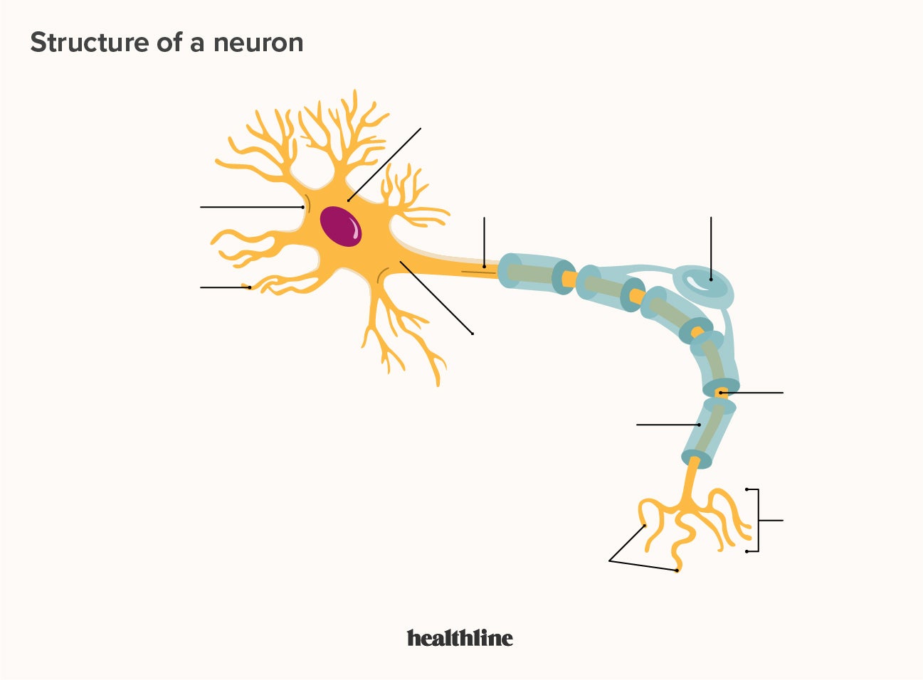
What Is a Neuron? Diagrams, Types, Function, and More
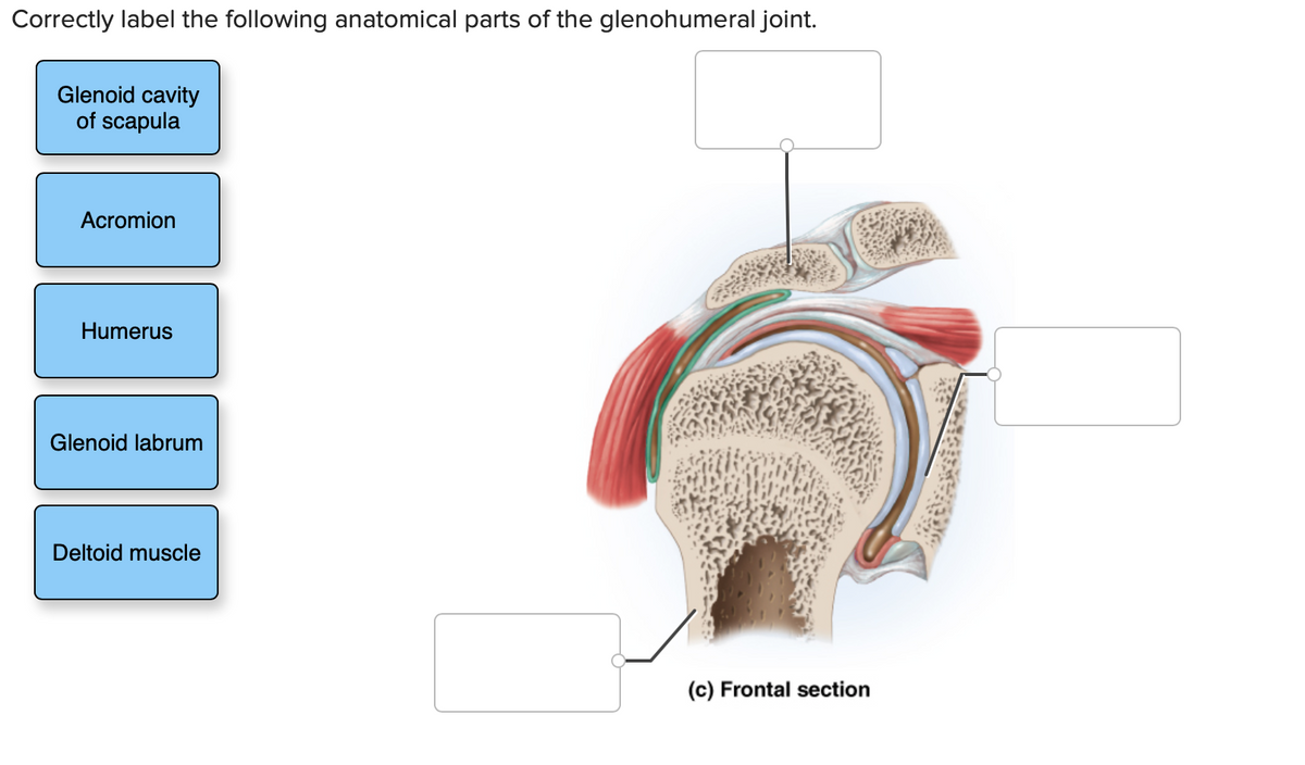
Answered: Glenoid cavity of scapula Acromion… | bartleby

Describe the structure and function of Neuron with labelled ...
Solved Help Correctly label the following anatomical | Chegg.com

Illustration of the anatomical features of the brain that are ...

Anatomy Exam 3 study guide Flashcards | Quizlet
Solved Correctly label the following anatomical features of ...
Chapter 12-Neural Tissue Flashcards - Easy Notecards

OneClass: What is the correct label of the following ...
Solved Saved Correctly label the following anatomical | Chegg.com
Post a Comment for "41 correctly label the following anatomical features of a neuron"